Pathogen Denial & Exosome Hypothesis of Viruses Debunked For Good
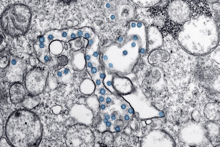
Transmission electron microscopic image of an isolate from the first U.S. case of COVID-19. The spherical viral particles, colourized blue, contain cross-sections through the viral genome, seen as black dots. (U.S. CDC)
WHAT ARE EXOSOMES?
I was just doing more reading on exosomes, for the debunking of the false "theory" going around that "viruses are only being confused with exosomes, and are not pathogenic" and wanted to illustrate how reading the science literature requires deep understanding of the mechanistic cellular processes, which can only be understood in reading by having foundational understanding of the terminology used to describe the mechanism of action. This is primal to how the study of language etymology is so fundamental to all forms of understanding in the fields of learning.
If you dont accurately understand the story the words are telling and why, you arrive at false conclusions.
Specialists in their field understand these mechanisms by training in that specific field and being taught what those terms mean as they go and are specific to their study, but a master etymologist could also accurately come to know the mechanisms of just about anything in the world by understanding the root meanings of the terminology building the way things work around us, and is used to understand HOW THINGS COME TO MEAN WHAT THEY DO and it deeply describes the characteristics and qualities of that which is being described.
When something new is discovered, the etymology is KEY in archiving what that new discovery means and how it will be subsequently transmitted.
To build a temple of wisdom, you must first place all the brick and mortar, exactly the way language paves the way with blocks to build a concept of understanding, a "structure to stand under."
Lets do a brief exercise in learning how sars cov2 novel coronavirus (covid19) is NOT being confused with exosomes of intracellular vesicular origin (the body's own native cellular shedding)
start with what exosomes actually are:
"Exosomes are best defined as extracellular vesicles that are released from cells upon fusion of an intermediate endocytic compartment, the multivesicular body (MVB), with the plasma membrane. This liberates intraluminal vesicles (ILVs) into the extracellular milieu and the vesicles thereby released are what we know as exosomes." https://bmcbiol.biomedcentral.com/articles/10.1186/s12915-016-0268-z
Now you're probably left wondering what all that sciencey jargon means, and are probably not any closer to fundamentally understanding what it is.
The fact is that those "sciencey words" are actually the most inexhaustive way to explain in the least amount of words and effort with detailed understanding, because each word has a whole process and story behind what it means.
Lets break it down:
exosome means "outside body" exo "outside", soma "body", referring to what is outside the cellular body, the cell boundary, so an exosome is a cellular body outside a cellular body.
These are called extracellular VESICLES meaning SACK OUTSIDE THE CELL, which can be understood by its etymology of VESICA "bladder" as a bladder means "inflated SACK," filled with fluid, as in VESICA PISCIS "fish bladder" which is also the geometric depiction of two cellular bodies (sacks) dividing or merging together, just as what happens when an intracellular (inside the cell), more descriptively an intraluminal space opens up as the cell takes something within it and encapsulates it in its own plasma membrane, basically a cell within a cell, the endocytic compartment forms, and either moves inward to the lysosome to degrade the intraluminal waste or it moves outward and buds or emerges out from inside the cell into the interstitial space (between the cells).
"In biology , a lumen (plural LUMINA) is the INSIDE SPACE of a tubular structure, such as an artery or intestine. [1]1 It comes from Latin lumen, meaning 'AN OPENING'."
So an INTRA (into) LUMINAL space is an "opening into" the cell.
This process is called ENDOCYTOSIS, and is defined as " a cellular process in which substances are brought into the cell. The material to be internalized is surrounded by an area of cell membrane, which then buds off inside the cell to form a vesicle containing the ingested material."
So a virus simply hijacks this already occurring process in the cell after successfully binding to the ACE-2 receptor protein on the surface of the cell membrane to be taken in through endocytosis.
This means upon replication of the virus within the cell, virions, little viral particles of dna or rna get released from the cell as exosomes. Virions are exosomes, but native cellular exosomes are NEVER virions.
There is literally no confusion about this process in cellular molecular biology or virology. The only confusion comes from the layman who doesnt understand this process and is not specializing in the field, such as pathogen deniers like Dr kaufman, Thomas Cowan, Andreas Vanderplanitz, Tom Barnett, and Sayerji.
Sayerji of Greenmedinfo: has ZERO QUALIFICATIONS OR EXPERTISE IN THE FIELD!
"INTRODUCTION TO VIROLOGY
Virology is the branch of microbiology that deals with the study of viruses and viral diseases in detail. Wendell Meredith Stanley ( 16 August 1904 – 15 June 1971 ) was an American biochemist, virologist and Nobel laureate Known as the Father of Virology.
GENERAL CHARACTERISTICS OF VIRUSES
⇒ Viruses are the organisms that separate living from non-living.
⇒ Cell organelles are absent in viruses.⇒ Viruses possess genetic material, either DNA or RNA.
⇒ Viruses lack enzymes except for HIV (Contains Reverse Transcriptase Enzyme).
⇒ Viruses are obligate intracellular parasites.
⇒ Viruses multiply by complex processes and not by binary fission.
⇒ They are unaffected by Antibiotics but affected by Interferon.
MORPHOLOGY OF VIRUSES
⇒ Most of the viruses are roughly spherical in shape.
⇒ Some viruses are irregular and pleomorphic
⇒ For e.g. –TOBACCO MOSAIC VIRUS – Rod shape RABIES VIRUS – Bullet Shape POX VIRUS – Brick shape
⇒ Viruses are measured in nanometers (nm)⇒ 1 nm = 10-9 m (0.000000001 m)
⇒ The largest virus is the Smallpox virus – 300 nm.
⇒ The smallest virus is the Parvovirus – 20 nm.
RIVER’S POSTULATES hese postulates were proposed by Thomas M. River in 1937 to establish the role of a specific virus as the cause of a specific disease. These postulates are the modifications of Koch’s postulates.
⇒ The viral agent must be found either in the host’s (animal or plant) body fluids at the time of disease or in cells showing lesions specific to that disease.
⇒ The host material with the viral agent used to inoculate the healthy host (test organism) must be free of any other microorganism.
⇒ The viral agent obtained from the infected host must –Produce the specific disease in a suitable healthy host,And/orProvide evidence of infection by inducing the formation of antibodies specific to that agent.
⇒ Similar material (viral particle) from the newly infected host (test organism) must be isolated and capable of transmitting the specific disease to other healthy hosts.
CLASSIFICATION OF VIRUSES
Viruses have been classified on the basis of Genetic Material & Symmetry: –
⇒ On the basis of Genetic Material
DNA virusesRNA virusesBoth DNA & RNA viruses are further classified as –i.) Single-Stranded DNA (ssDNA) or Double-Stranded DNA (dsDNA) viruses.ii.) Single-Stranded RNA (ssRNA) or Double-Stranded RNA (dsRNA) viruses.
⇒ On the basis of Symmetry
IcosahedralHelicalComplex
An Icosahedral Symmetry is described as a polygon with 12 vertices or corners and 20 facets in the shape of equilateral triangular faces. E.g. – Papovavirus, Picornavirus, Herpes virus, Adenoviruses etc.
In Helical Symmetry, the nucleic acid and capsomeres are wound together to form a helical or spiral tube. Most of the helical viruses are enveloped & all are RNA viruses. E.g. – Tobacco mosaic virus (TMV), influenza virus etc.
Those viruses which have more complex structure and do not fit into either category (Icosahedral or Helical) are placed under Complex Symmetry. g. – Poxviruses and Large Bacteriophages etc.
The outbreak sent researchers around the world racing to isolate laboratory specimens of the virus that causes COVID-19. The virus was later named severe acute respiratory syndrome coronavirus 2, or SARS-CoV-2.In countries that experienced earlier outbreaks, including China, Australia, Germany and the United States, researchers were able to isolate the virus and develop their own inventories of SARS-CoV-2, but logistical and legal barriers prevented them from readily sharing their materials with researchers beyond their borders.What Canadian researchers needed to join the fight in earnest was a domestic supply of clean copies of the virus — preferably from multiple Canadian COVID-19 cases.
Arinjay Banerjee, a postdoctoral research fellow at McMaster who typically works in my virology lab, volunteered his special expertise. We were proud to have him share his talent with the team in Toronto, where he set to work with physicians and researchers Samira Mubareka, Lily Yip, Patryk Aftanas and Rob Kozak.For Banerjee, it was like a batter being called to the plate with the score tied in the bottom of the ninth.
Ideal viral conditionsIsolating a virus requires collecting specimens from patients and culturing, or growing, any viruses that occur in the samples. These viruses are obligate intracellular parasites, which means that they can only replicate and multiply in cells. To isolate a particular virus, researchers need to provide it with an opportunity to infect live mammalian cells, in tiny flasks or on tissue culture plates.Viruses adapt to their hosts and evolve to survive and replicate efficiently within their particular environment. When a new virus such as SARS-CoV-2 emerges, it isn’t obvious what particular environment that virus has adapted to, so it can be hard to grow it successfully in the lab.We can use tricks to draw out a virus. Sometimes the tricks work and sometimes they don’t.
In this case, the researchers tried a method Banerjee and the team had previously used while working on the coronavirus that causes Middle Eastern Respiratory Syndrome: culturing the virus on immunodeficient cells that would allow the virus to multiply unchecked. It worked. Since specimens from patients are also likely to contain other viruses, it is critical to determine if a virus growing in the culture is really the target coronavirus. Researchers confirm the source of infection by extracting genetic material from the virus in culture and sequencing its genome.They compare the sequence to known coronavirus sequences to identify it precisely. Once a culture is confirmed, researchers can make copies to share with colleagues.All this work must be done in secure, high-containment laboratories that mitigate the risk of accidental virus release into the environment and also protect scientists from accidental exposure.
The more versions of a virus that can be isolated, the better. Having multiple virus isolates allows us to monitor how the virus is evolving in humans as the pandemic progresses. It also allows researchers to test the efficacy of vaccines and drugs against multiple mutations of the virus.
The pathogen deniers go on to try to dismiss the research as false because of "funding tho" by making spurious connections to the Bill & Melinda Gates foundation to the University which the author is Vice President.
Maybe you should dig deeper and with less bias. The Gates foundation hasnt even donated to mcmaster since 2017.
The idiots like christine massey trying to claim that bill gates funded and conducted the canadian isolation studies is ridiculous and doesnt even follow sound research or logic.
just because gates foundation donated some money to mcmaster university for a few years until 2017...
"to identify and address ethical challenges, ethics-related risk, and policy gaps that have the potential to undermine the impact of potential life-saving technologies and interventions in global health and development research; Aid type: Project-type interventions. Affected regions: Developing countries, unspecified."
...and not in 2020 does not mean the author of the article and study, Karen Mossman, who is a researcher and vice president of mcmaster university, recieved funding direct funding from bill gates to conduct and design a fake study, and even if he DID directly fund her specifically, which he did not, that still would not discredit or invalidate the scientific findings of viral isolation in canada.
This whole nonsense about needing to fulfill "Koch's postulates" WHICH IS FOR BACTERIA, NOT VIRUSES, in order to know what SARS COV 2 is, how it works, what its protein spike structure is, the amino acid sequence and genome to differentiate it from a native cellular exosome is a ridiculous strawman argument.
Extracellular vesicles dont have s protein spikes that could in any way be confused with SARS COV. They dont have protein spikes at all. even a layperson can distinguish the difference in a monolayer culture by looking at them. its fun to learn about, yes, but it doesnt debunk viruses, especially not the entire field.
And no "pleomorphism" literally has no bearing on this research.
Pleomorphism doesnt apply to all microbes as highly unqualified Robert o Young arbitrarily claims. only certain microbes are pleomorphic and he doesnt have the evidence to claim otherwise. Sure speculating can be fun, but it doesnt discredit the entire field.
I assure you, if it was that big of a hoax on the micro level, there would be more than just Lanka talking about it. I assure you, publishing scientists in the field like Alina Chan, who publishes papers falsifying the "zoonotic origins" of it , would be saying it hasnt even been isolated, but they are not, because it has. The skeptics claiming it hasnt are not correct nor qualified to even know the difference.
Koch's postulates HAVE BEEN FULFILLED for SARS COV 2003.
"According to Koch's postulates, as modified by Rivers for viral diseases, six criteria are required to establish a virus as the cause of a disease1. The first three criteria — isolation of virus from diseased hosts, cultivation in host cells, and proof of filterability — have been met for SCV by several groups2,3,4,5. Moreover, of 96 individuals complying with the World Health Organization's definition of SARS6 in Hong Kong, 86 (90%) yielded laboratory evidence of SCV infection.We have tested for the three remaining criteria: production of comparable disease in the original host species or a related one, re-isolation of the virus, and detection of a specific immune response to the virus.
We inoculated two macaques with Vero-cell-cultured SCV isolated from a fatal SARS case, and monitored their clinical signs, virus excretion and antibody response. The animals were killed six days post-inoculation (d.p.i.), and we then carried out gross and histopathological examinations of them.Both SCV-inoculated macaques became lethargic from 3 d.p.i. onwards and developed a temporary skin rash, and one suffered respiratory distress from 4 d.p.i. onwards. The macaques excreted virus from the nose and throat at 2–6 d.p.i., as shown by polymerase chain reaction with reverse transcription (RT-PCR) and by virus isolation (see supplementary information).
The isolated virus was identical to that inoculated, as shown by negative-contrast electron microscopy (Fig. 1a) and RT-PCR analysis. Seroconversion to SCV, as determined by indirect immunofluorescence assay using infected Vero cells, was demonstrated in two other SCV-infected macaques at 16 d.p.i.. The virus was also isolated from the faeces of one of these animals (see supplementary information).
"An acute and often severe respiratory illness emerged in southern China in late 2002 and rapidly spread to different areas of the Far East as well as several countries around the globe. When the outbreak of this apparently novel infectious disease termed severe acute respiratory syndrome (SARS) came to an end in July 2003, it had caused over 8000 probable cases worldwide and more than 700 deaths.Starting in March 2003, the World Health Organization (WHO) organised an unprecedented international effort by leading laboratories working together to find the causative agent. Little more than one week later, three research groups from this WHO-coordinated network simultaneously found evidence of a hitherto unknown coronavirus in SARS patients, using different approaches.
After Koch’s postulates had been fulfilled, WHO officially declared on 16 April 2003 that this virus never before seen in humans is the cause of SARS.Ever since, progress around SARS-associated coronavirus (SARS-CoV) has been swift. Within weeks of the first isolate being obtained, its complete genome was sequenced. Diagnostic tests based on the detection of SARS-CoV RNA were developed and made available freely and widely; nevertheless the SARS case definition still remains based on clinical and epidemiological criteria"
"SARS-CoV-2 was first isolated using human airway epithelial cells and it was classified into the subgenus sarbecovirus of beta-CoVs by phylogenetic analyses of the gene sequences.3
Both the SARS-CoV and the MERS-CoV were initially isolated and grew readily in Vero cells.4, 5 Here, we report the isolation of SARS-CoV-2 using Vero cells from a patient entering Korea from Wuhan, China."
"Virus isolation from the clinical specimens was performed with human airway epithelial cells and Vero E6 and Huh-7 cell lines. The isolated virus was named 2019-nCoV."
"Although our study does not fulfill Koch’s postulates, our analyses provide evidence implicating 2019-nCoV in the Wuhan outbreak.
Additional evidence to confirm the etiologic significance of 2019-nCoV in the Wuhan outbreak include identification of a 2019-nCoV antigen in the lung tissue of patients by immunohistochemical analysis, detection of IgM and IgG antiviral antibodies in the serum samples from a patient at two time points to demonstrate seroconversion, and animal (monkey) experiments to provide evidence of pathogenicity. "
"According to Koch's postulates, as modified by Rivers for viral diseases, six criteria are required to establish a virus as the cause of a disease1.
The first three criteria — isolation of virus from diseased hosts, cultivation in host cells, and proof of filterability — have been met for SCV by several groups2,3,4,5.
Moreover, of 96 individuals complying with the World Health Organization's definition of SARS6 in Hong Kong, 86 (90%) yielded laboratory evidence of SCV infection.
We have tested for the three remaining criteria: production of comparable disease in the original host species or a related one, re-isolation of the virus, and detection of a specific immune response to the virus. We inoculated two macaques with Vero-cell-cultured SCV isolated from a fatal SARS case, and monitored their clinical signs, virus excretion and antibody response. The animals were killed six days post-inoculation (d.p.i.), and we then carried out gross and histopathological examinations of them.Both SCV-inoculated macaques became lethargic from 3 d.p.i. onwards and developed a temporary skin rash, and one suffered respiratory distress from 4 d.p.i. onwards. The macaques excreted virus from the nose and throat at 2–6 d.p.i., as shown by polymerase chain reaction with reverse transcription (RT-PCR) and by virus isolation (see supplementary information).
The isolated virus was identical to that inoculated, as shown by negative-contrast electron microscopy (Fig. 1a) and RT-PCR analysis.
Seroconversion to SCV, as determined by indirect immunofluorescence assay using infected Vero cells, was demonstrated in two other SCV-infected macaques at 16 d.p.i..
The pathogen deniers only arbitrary rebuttal to dismiss all the studies is that one study used fetal bovine serum as viral transport medium to isolate and purify sars cov 2 and that "contaminates the sample with bovine rna which is somehow confused with sars cov rna gene sequence.
They just simply dismiss and deny all evidence because they falsely assume that the methodologies used to culture and isolate and purify viral samples, are somehow "contaminated with antibiotics, antiviral and toxic agents".
He goes on to suggest that "antifungals and fetal bovine serum and fetal bovine serum can interfere with the RNA in the sample," based on the cherry picked misinterpretation and misunderstanding of the citation.
""The authors first evaluated exogenous RNA contamination. They grew cultures of a cell type known not to express a particular RNA, then evaluated the presence of that RNA in the culture media. If that RNA was found, its origin was probably the media itself. For example, the authors demonstrated that miR-122, a liver-specific miRNA, is present in media from cultured glioma cells, suggesting that its source is likely FBS itself. They then attempted to deplete RNA from FBS via ultracentrifugation, but despite a 24 hour spin at 100,000g, about 75% of total RNA remained in the supernatant. This result has also been found by researchers attempting to deplete FBS of RNA-containing EVs and emphasizes the difficulty of producing media truly free from contaminating RNA."
But he leaves out the other part that suggests they can distinguish between which rna genome is from a virus like sars cov 2 rather than fetal bovine serum.
"Alternatively, a quantitative analysis of the chemical composition of the media might make it possible to estimate which RNAs are secreted by the cells of interest by filtering out known FBS RNAs from the total RNA pool."
can you please read what sterile and heat inactivated means please?
Heat inactivated bovine serum is heat inactivated to sterilize it.
Heat inactivation of serum refers to the process which involves treatment of serum at a higher temperature to inactivate unwanted active agents in serum particularly serum complement.
In addition to inhibiting the complement system, heat inactivation is also reported to reduce the risk of microbial contaminants like mycoplasma. However, heat inactivation often results in precipitation, and serum appears translucent. Precipitates in serum are frequently mistaken for microbial contaminants.
"Heat treatment was also advocated in the past as a risk mitigation approach for achieving pathogen (especially mycoplasma and virus) reduction in serum. "
"It has been showniii that mycoplasma and certain viruses (including such known contaminants of bovine serum as bovine viral diarrhea virus, parainfluenza type 3, infectious bovine rhinotracheitis, reovirus type 3, and adenovirus) are rendered essentially non-infectious at the temperatures/treatment times used for heat-inactivation.
There are, however, other ways of inactivating such potential contaminants that are less potentially damaging to the serum (see our article on gamma irradiation of serum for risk mitigation, for instance). "
"Animal-derived materials such as animal sera represent a low, but finite, risk for introduction of an adventitious agent (virus or mollicute) into a biological bulk harvest during upstream manufacturing processes involving mammalian cell substrates.Viral and mollicute (Mycoplasma sp. and Acholeplasma sp.) contamination events have been relatively rare, but many of those that have been reported have been attributed to use of infected animal sera in growth media during cell expansion.The risk of introduction of viruses and mollicutes may be mitigated by elimination of the use of animal sera and implementation instead of chemically defined or serum- and animal-derived material-free cell culture media.When use of animal sera is unavoidable, however, mitigation of the risk of introducing an adventitious contaminant may involve treatment of the sera to inactivate potential contaminants. Gamma irradiation is one of the most widely employed methods for viral and mollicute inactivation in animal sera.In this article, we review the inactivation results reported for viral and mollicute inactivation in frozen serum. Studies performed to assess the impact of gamma irradiation on serum quality and performance are also discussed.The available data indicate that inactivation of mollicutes in serum is essentially complete at the gamma radiation doses normally employed (25–40 kGy),
Isolation and rapid sharing of the 2019 novel coronavirus (SARS-CoV-2) from the first patient diagnosed with COVID-19 in Australia https://pubmed.ncbi.nlm.nih.gov/32237278/
Scientists figure out how new coronavirus breaks into human cells (endocytosis):
"Zhou and his team used a tool called cryo electron microscopy, which employs deeply frozen samples and electron beams to image the tiniest structures of biological molecules. The researchers found that the molecular bond between SARS-CoV-2's spike protein and ACE2 looks fairly similar to the binding pattern of the coronavirus that caused the outbreak of SARS in 2003.
There are some differences, however, in the precise amino acids used to bind SARS-CoV-2 to that ACE2 receptor compared with the virus that causes SARS (severe acute respiratory syndrome), the researchers said. "While some might consider the differences subtle," Gallagher said, "they might be meaningful with respect to the strength with which each of those viruses stick." That "stickiness" could affect how easily a virus transmits from one person to another. If any given viral particle is more likely to enter a cell once it enters the human body, transmission of disease is more likely.There are other coronaviruses that circulate regularly, causing upper respiratory infections that most people think of as the common cold. Those coronaviruses don't interact with the ACE2 receptor, Gallagher said, but rather, they get into the body using other receptors on human cells. " https://www.livescience.com/how-coronavirus-infects-cells.html
Structural basis for the recognition of SARS-CoV-2 by full-length human ACE2
Abstract
"Angiotensin-converting enzyme 2 (ACE2) is the cellular receptor for severe acute respiratory syndrome–coronavirus (SARS-CoV) and the new coronavirus (SARS-CoV-2) that is causing the serious coronavirus disease 2019 (COVID-19) epidemic. Here, we present cryo–electron microscopy structures of full-length human ACE2 in the presence of the neutral amino acid transporter B0AT1 with or without the receptor binding domain (RBD) of the surface spike glycoprotein (S protein) of SARS-CoV-2, both at an overall resolution of 2.9 angstroms, with a local resolution of 3.5 angstroms at the ACE2-RBD interface. The ACE2-B0AT1 complex is assembled as a dimer of heterodimers, with the collectrin-like domain of ACE2 mediating homodimerization. The RBD is recognized by the extracellular peptidase domain of ACE2 mainly through polar residues. These findings provide important insights into the molecular basis for coronavirus recognition and infection."
"SARS-CoV transmission electron microscopy. In the supernatant of SARS-CoV infected cytopathic Vero E6 cells, characteristic virus particles can be found. The diameter of the viruses ranges between 60 nm and 120 nm and the virus shapes are round or oval. There are many protrusions from the envelope which are arranged in order with wide gaps between them. There are also many virus particles in the infected cells present. They often form a virus vesicle with an encircling membrane. A: Higher magnification B: Lower magnification. Scale bars represent 100 nm. Reproduced with permission from Acta Biochimica et Biophysica Sinica 2003, 35(6):587–591 [126]."
Clarifying the statement provided by FDA in the SARS CoV2 PCR method document.
"Since no quantified virus isolates of the 2019-nCoV are currently available, assays designed for detection of the 2019-nCoV RNA were tested with characterized stocks of in vitro transcribed full length RNA (N gene; GenBank accession: MN908947.2) of known titer (RNA copies/μL) spiked into a diluent consisting of a suspension of human A549 cells and viral transport medium (VTM) to mimic clinical specimen”
One essential step in method characterization is to determine the limit of detection (LoD). As this requires a quantitative readout from the assay, one needs to use a precisely quantified reference substance as calibrant, ie it needs to be known in the reference that the SARS CoV2 RNA genome is exactly present at a concentration of X copies/µl.
This statement simply means that this exactly quantified isolate is not availabe, but the isolate are available. The exactly quantified reference substance is available in the form of in-vitro transcribed RNA. This was used for the quantitative studies to establish the LoD.
This statement does NOT mean that no isolates are available. The CDC distributes currently 19 different SARS CoV2 isolates.
A very simple analogy:
apples = SARS CoV2 isolates
Do you have apples available?
Yes
Do you have buckets of exactly 50 apples available?
No
But you do have apples in store?
Yes, we do and we sell them in buckets, but some buckets contain as little as 40 apples and some may contain up to 60 apples.“
Make sure to like, subscribe and hit the bell notifications button for full films and more videos! access my archives @
Show your support by giving the gift of knowledge and wisdom from the Official Lifting The Veil Merch shop for epic and inspiring apparel and art designs!
All of my full video presentations and articles can be found at SUBSCRIBESTAR.COM/LIFTING-THE-VEILfor full #esoteric film presentations, research articles, livestream hangouts, lessons and so much more mind blowing esoteric content on theology, mythology, etymology and language, symbolism, science, biology, health and so much more!
SEE YOU SOON!
MUCH LOVE, PEACE AND WISDOM!
------------SUPPORT Lifting The veil -------------------
SubscribeStar and its trusted third parties collect browsing information as specified in the Privacy Policy and use cookies or similar technologies for analysis and technical purposes and, with your consent, for functionality, experience, and measurement as specified in the Cookies Policy.