Join for free to receive email notifications about new posts submitted to paid tiers
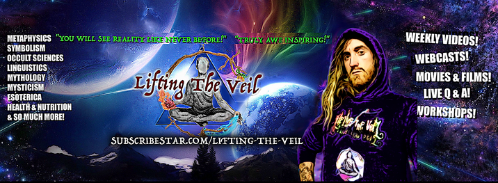
Thanks so much for your support! Let me know I can best Serve you! GIve your feedback!
Recieve Credits for making production possible in all my films
Get access to live voice and text chat on air during Lifting The Veil Live broadcasts!
As a patron, you'll be recognized for making it possible in all my full video presentations.
Get access to live voice and text chat on air during Lifting The Veil Live broadcasts! Credits on an upcoming video Plus all previous rewards
Get access to live voice and text chat on air during Lifting The Veil Live broadcasts!
Get full access to what im working on as well as my extended cuts. All Rewards in lower Tiers
free copy of my E-book article "CALLING ALL RELIGIONS! TRUE ANCIENT HIDDEN EVIDENCE OF RELIGIOUS ORIGIN!" (upon request)
Free Ebook copies of all of my presentation archives inluding unfinished draft projects before publication! (upon request) DVD or Blu Ray HD copies of any of my full films and videos of your choice! PDF ebooks of all of my presentations! (Upon Request) weekly Q&A hangout to discuss anything you want!
All perks of lower tiers upon request!
+Free access to full one hour skype session per week by request for absolutely anything you want to talk about
All the rewards in Lower Tiers,
"Exosomes are best defined as extracellular vesicles that are released from cells upon fusion of an intermediate endocytic compartment, the multivesicular body (MVB), with the plasma membrane. This liberates intraluminal vesicles (ILVs) into the extracellular milieu and the vesicles thereby released are what we know as exosomes." https://bmcbiol.biomedcentral.com/articles/10.1186/s12915-016-0268-z
"INTRODUCTION TO VIROLOGY
Viral isolation
The outbreak sent researchers around the world racing to isolate laboratory specimens of the virus that causes COVID-19. The virus was later named severe acute respiratory syndrome coronavirus 2, or SARS-CoV-2.In countries that experienced earlier outbreaks, including China, Australia, Germany and the United States, researchers were able to isolate the virus and develop their own inventories of SARS-CoV-2, but logistical and legal barriers prevented them from readily sharing their materials with researchers beyond their borders.What Canadian researchers needed to join the fight in earnest was a domestic supply of clean copies of the virus — preferably from multiple Canadian COVID-19 cases.
Even in a pandemic, developing such a supply is not as easy as it might sound, and multiple teams in Canada set out to isolate and develop pure cultures of the virus, not knowing which would be successful, or when.Ultimately two teams in Canada would isolate the virus for study: one at the University of Saskatchewan and one that featured researchers from McMaster University, Sunnybrook Health Sciences Centre and the University of Toronto.
Arinjay Banerjee, a postdoctoral research fellow at McMaster who typically works in my virology lab, volunteered his special expertise. We were proud to have him share his talent with the team in Toronto, where he set to work with physicians and researchers Samira Mubareka, Lily Yip, Patryk Aftanas and Rob Kozak.For Banerjee, it was like a batter being called to the plate with the score tied in the bottom of the ninth.
He had come to work at McMaster because of its Institute for Infectious Disease Researchand its Immunology Research Centre, and because the university maintains a research colony of bats.Banerjee’s PhD work at the University of Saskatchewan, and now at McMaster, has focused on bats and how their viruses, including coronaviruses, interact with bat and human antiviral responses. Over the past few years, studies have shown that bat coronaviruses have the capacity to infect human cells. Multiple researchers had predicted a coronavirus that would evolve and jump into humans.
Read more: It's wrong to blame bats for the coronavirus epidemic
Ideal viral conditionsIsolating a virus requires collecting specimens from patients and culturing, or growing, any viruses that occur in the samples. These viruses are obligate intracellular parasites, which means that they can only replicate and multiply in cells. To isolate a particular virus, researchers need to provide it with an opportunity to infect live mammalian cells, in tiny flasks or on tissue culture plates.Viruses adapt to their hosts and evolve to survive and replicate efficiently within their particular environment. When a new virus such as SARS-CoV-2 emerges, it isn’t obvious what particular environment that virus has adapted to, so it can be hard to grow it successfully in the lab.We can use tricks to draw out a virus. Sometimes the tricks work and sometimes they don’t.
In this case, the researchers tried a method Banerjee and the team had previously used while working on the coronavirus that causes Middle Eastern Respiratory Syndrome: culturing the virus on immunodeficient cells that would allow the virus to multiply unchecked. It worked. Since specimens from patients are also likely to contain other viruses, it is critical to determine if a virus growing in the culture is really the target coronavirus. Researchers confirm the source of infection by extracting genetic material from the virus in culture and sequencing its genome.They compare the sequence to known coronavirus sequences to identify it precisely. Once a culture is confirmed, researchers can make copies to share with colleagues.All this work must be done in secure, high-containment laboratories that mitigate the risk of accidental virus release into the environment and also protect scientists from accidental exposure.
The more versions of a virus that can be isolated, the better. Having multiple virus isolates allows us to monitor how the virus is evolving in humans as the pandemic progresses. It also allows researchers to test the efficacy of vaccines and drugs against multiple mutations of the virus.
Canadian viral strains
"to identify and address ethical challenges, ethics-related risk, and policy gaps that have the potential to undermine the impact of potential life-saving technologies and interventions in global health and development research; Aid type: Project-type interventions. Affected regions: Developing countries, unspecified."
https://donations.vipulnaik.com/donee.php?donee=McMaster%20University&fbclid=IwAR3WNwKm2vRM_ljAZdk5SaFRRo_YIH7NkzLyuJ5gPCtL5Wkqlt3EZoV8qIo#doneeDonationAmountsByDonorAndYear
"According to Koch's postulates, as modified by Rivers for viral diseases, six criteria are required to establish a virus as the cause of a disease1. The first three criteria — isolation of virus from diseased hosts, cultivation in host cells, and proof of filterability — have been met for SCV by several groups2,3,4,5. Moreover, of 96 individuals complying with the World Health Organization's definition of SARS6 in Hong Kong, 86 (90%) yielded laboratory evidence of SCV infection.We have tested for the three remaining criteria: production of comparable disease in the original host species or a related one, re-isolation of the virus, and detection of a specific immune response to the virus.
We inoculated two macaques with Vero-cell-cultured SCV isolated from a fatal SARS case, and monitored their clinical signs, virus excretion and antibody response. The animals were killed six days post-inoculation (d.p.i.), and we then carried out gross and histopathological examinations of them.Both SCV-inoculated macaques became lethargic from 3 d.p.i. onwards and developed a temporary skin rash, and one suffered respiratory distress from 4 d.p.i. onwards. The macaques excreted virus from the nose and throat at 2–6 d.p.i., as shown by polymerase chain reaction with reverse transcription (RT-PCR) and by virus isolation (see supplementary information).
The isolated virus was identical to that inoculated, as shown by negative-contrast electron microscopy (Fig. 1a) and RT-PCR analysis. Seroconversion to SCV, as determined by indirect immunofluorescence assay using infected Vero cells, was demonstrated in two other SCV-infected macaques at 16 d.p.i.. The virus was also isolated from the faeces of one of these animals (see supplementary information).
"An acute and often severe respiratory illness emerged in southern China in late 2002 and rapidly spread to different areas of the Far East as well as several countries around the globe. When the outbreak of this apparently novel infectious disease termed severe acute respiratory syndrome (SARS) came to an end in July 2003, it had caused over 8000 probable cases worldwide and more than 700 deaths.Starting in March 2003, the World Health Organization (WHO) organised an unprecedented international effort by leading laboratories working together to find the causative agent. Little more than one week later, three research groups from this WHO-coordinated network simultaneously found evidence of a hitherto unknown coronavirus in SARS patients, using different approaches.
After Koch’s postulates had been fulfilled, WHO officially declared on 16 April 2003 that this virus never before seen in humans is the cause of SARS.Ever since, progress around SARS-associated coronavirus (SARS-CoV) has been swift. Within weeks of the first isolate being obtained, its complete genome was sequenced. Diagnostic tests based on the detection of SARS-CoV RNA were developed and made available freely and widely; nevertheless the SARS case definition still remains based on clinical and epidemiological criteria"
We have tested for the three remaining criteria: production of comparable disease in the original host species or a related one, re-isolation of the virus, and detection of a specific immune response to the virus. We inoculated two macaques with Vero-cell-cultured SCV isolated from a fatal SARS case, and monitored their clinical signs, virus excretion and antibody response. The animals were killed six days post-inoculation (d.p.i.), and we then carried out gross and histopathological examinations of them.Both SCV-inoculated macaques became lethargic from 3 d.p.i. onwards and developed a temporary skin rash, and one suffered respiratory distress from 4 d.p.i. onwards. The macaques excreted virus from the nose and throat at 2–6 d.p.i., as shown by polymerase chain reaction with reverse transcription (RT-PCR) and by virus isolation (see supplementary information).
The isolated virus was identical to that inoculated, as shown by negative-contrast electron microscopy (Fig. 1a) and RT-PCR analysis.
Seroconversion to SCV, as determined by indirect immunofluorescence assay using infected Vero cells, was demonstrated in two other SCV-infected macaques at 16 d.p.i..
The virus was also isolated from the faeces of one of these animals (see supplementary information). https://academic.oup.com/cid/article/doi/10.1093/cid/ciaa325/5811871
""The authors first evaluated exogenous RNA contamination. They grew cultures of a cell type known not to express a particular RNA, then evaluated the presence of that RNA in the culture media. If that RNA was found, its origin was probably the media itself. For example, the authors demonstrated that miR-122, a liver-specific miRNA, is present in media from cultured glioma cells, suggesting that its source is likely FBS itself. They then attempted to deplete RNA from FBS via ultracentrifugation, but despite a 24 hour spin at 100,000g, about 75% of total RNA remained in the supernatant. This result has also been found by researchers attempting to deplete FBS of RNA-containing EVs and emphasizes the difficulty of producing media truly free from contaminating RNA."
https://www.cdc.gov/coronavirus/2019-ncov/downloads/Viral-Transport-Medium.pdf?fbclid=IwAR04CAMGDlOWiweSVGbZHogl6PGScw8ellxfq4PaolPN_Kk4zBC2C0vgc_0
"Animal-derived materials such as animal sera represent a low, but finite, risk for introduction of an adventitious agent (virus or mollicute) into a biological bulk harvest during upstream manufacturing processes involving mammalian cell substrates.Viral and mollicute (Mycoplasma sp. and Acholeplasma sp.) contamination events have been relatively rare, but many of those that have been reported have been attributed to use of infected animal sera in growth media during cell expansion.The risk of introduction of viruses and mollicutes may be mitigated by elimination of the use of animal sera and implementation instead of chemically defined or serum- and animal-derived material-free cell culture media.When use of animal sera is unavoidable, however, mitigation of the risk of introducing an adventitious contaminant may involve treatment of the sera to inactivate potential contaminants. Gamma irradiation is one of the most widely employed methods for viral and mollicute inactivation in animal sera.In this article, we review the inactivation results reported for viral and mollicute inactivation in frozen serum. Studies performed to assess the impact of gamma irradiation on serum quality and performance are also discussed.The available data indicate that inactivation of mollicutes in serum is essentially complete at the gamma radiation doses normally employed (25–40 kGy),
"Zhou and his team used a tool called cryo electron microscopy, which employs deeply frozen samples and electron beams to image the tiniest structures of biological molecules. The researchers found that the molecular bond between SARS-CoV-2's spike protein and ACE2 looks fairly similar to the binding pattern of the coronavirus that caused the outbreak of SARS in 2003.
There are some differences, however, in the precise amino acids used to bind SARS-CoV-2 to that ACE2 receptor compared with the virus that causes SARS (severe acute respiratory syndrome), the researchers said. "While some might consider the differences subtle," Gallagher said, "they might be meaningful with respect to the strength with which each of those viruses stick." That "stickiness" could affect how easily a virus transmits from one person to another. If any given viral particle is more likely to enter a cell once it enters the human body, transmission of disease is more likely.There are other coronaviruses that circulate regularly, causing upper respiratory infections that most people think of as the common cold. Those coronaviruses don't interact with the ACE2 receptor, Gallagher said, but rather, they get into the body using other receptors on human cells. " https://www.livescience.com/how-coronavirus-infects-cells.html
"Angiotensin-converting enzyme 2 (ACE2) is the cellular receptor for severe acute respiratory syndrome–coronavirus (SARS-CoV) and the new coronavirus (SARS-CoV-2) that is causing the serious coronavirus disease 2019 (COVID-19) epidemic. Here, we present cryo–electron microscopy structures of full-length human ACE2 in the presence of the neutral amino acid transporter B0AT1 with or without the receptor binding domain (RBD) of the surface spike glycoprotein (S protein) of SARS-CoV-2, both at an overall resolution of 2.9 angstroms, with a local resolution of 3.5 angstroms at the ACE2-RBD interface. The ACE2-B0AT1 complex is assembled as a dimer of heterodimers, with the collectrin-like domain of ACE2 mediating homodimerization. The RBD is recognized by the extracellular peptidase domain of ACE2 mainly through polar residues. These findings provide important insights into the molecular basis for coronavirus recognition and infection."
"SARS-CoV transmission electron microscopy. In the supernatant of SARS-CoV infected cytopathic Vero E6 cells, characteristic virus particles can be found. The diameter of the viruses ranges between 60 nm and 120 nm and the virus shapes are round or oval. There are many protrusions from the envelope which are arranged in order with wide gaps between them. There are also many virus particles in the infected cells present. They often form a virus vesicle with an encircling membrane. A: Higher magnification B: Lower magnification. Scale bars represent 100 nm. Reproduced with permission from Acta Biochimica et Biophysica Sinica 2003, 35(6):587–591 [126]."
Explained here (& see pics
“Dear Editor,
Responding to Bernadette Pajer and Janet Menage.
Clarifying the statement provided by FDA in the SARS CoV2 PCR method document.
"Since no quantified virus isolates of the 2019-nCoV are currently available, assays designed for detection of the 2019-nCoV RNA were tested with characterized stocks of in vitro transcribed full length RNA (N gene; GenBank accession: MN908947.2) of known titer (RNA copies/μL) spiked into a diluent consisting of a suspension of human A549 cells and viral transport medium (VTM) to mimic clinical specimen”
One essential step in method characterization is to determine the limit of detection (LoD). As this requires a quantitative readout from the assay, one needs to use a precisely quantified reference substance as calibrant, ie it needs to be known in the reference that the SARS CoV2 RNA genome is exactly present at a concentration of X copies/µl.
This statement simply means that this exactly quantified isolate is not availabe, but the isolate are available. The exactly quantified reference substance is available in the form of in-vitro transcribed RNA. This was used for the quantitative studies to establish the LoD.
This statement does NOT mean that no isolates are available. The CDC distributes currently 19 different SARS CoV2 isolates.
A very simple analogy:
apples = SARS CoV2 isolates
Do you have apples available?
Yes
Do you have buckets of exactly 50 apples available?
No
But you do have apples in store?
Yes, we do and we sell them in buckets, but some buckets contain as little as 40 apples and some may contain up to 60 apples.“